Down-regulating GSK3β alleviates hypoxia/reoxygenation-induced injury of senescent renal tubular epithelial cells by inhibiting the function of ITPR1-GRP75-VDAC1 complex
-
摘要:
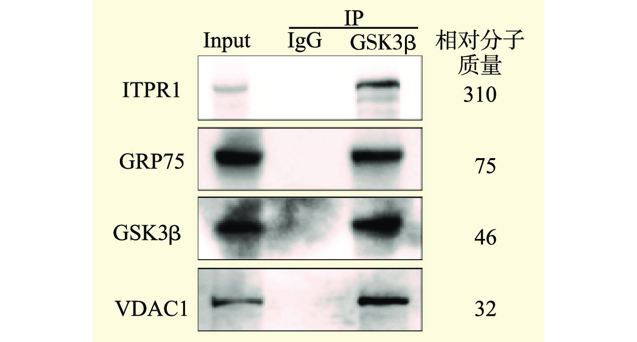
目的 探讨糖原合成酶激酶3β(GSK3β)对衰老小鼠原代肾小管上皮细胞(RTEC)缺氧/复氧(H/R)损伤的影响及其调控机制。 方法 将RTEC分成为Young组即正常生长的年轻RTEC、Old组即使用Etoposide诱导的衰老RTEC、Old+Ad-shNC+H/R组即使用Etoposide诱导衰老再转染腺病毒阴性对照(Ad-shNC)后进行H/R处理,Old+Ad-shGSK3β+H/R组即使用Etoposide诱导衰老后再转染靶向沉默GSK3β的短发夹RNA腺病毒(Ad-shGSK3β)后进行H/R处理。采用流式细胞术检测各组细胞凋亡水平和线粒体活性氧水平,采用免疫荧光染色法检测各组钙离子水平,采用蛋白质印迹法检测各组GSK3β、线粒体相关的内质网膜结构(MAM)相关蛋白肌醇1,4,5-三磷酸受体1(ITPR1)、电压依赖性阴离子通道1(VDAC1)、葡萄糖调节蛋白75(GRP75)表达及磷酸化水平,采用免疫共沉淀分析GSK3β与MAM相关蛋白的相互作用。 结果 与Young组比较,Old组细胞凋亡水平、线粒体活性氧水平及线粒体钙离子水平均较高;与Old组相比,Old+Ad-shNC+H/R组细胞凋亡水平、线粒体活性氧水平及线粒体钙离子水平均较高;与Old+Ad-shNC+H/R组比较,Old+Ad-shGSK3β+H/R组细胞凋亡水平、线粒体活性氧水平及线粒体钙离子水平均较低,差异均有统计学意义(均为P<0.05)。与Young组比较,Old组ITPR1、GRP75和GSK3β总蛋白表达增多,ITPR1和GRP75磷酸化水平升高,而VDAC1总蛋白和磷酸化蛋白水平均下降;与Old组比较,Old+Ad-shNC+H/R组GSK3β蛋白表达不变,ITPR1和GRP75总蛋白和磷酸化水平升高,VDAC1总蛋白表达不变,磷酸化水平增高;与Old+Ad-shNC+H/R组比较,Old+Ad-shGSK3β+H/R组GSK3β蛋白表达减少,ITPR1、GRP75和VDAC1总蛋白表达不变,磷酸化水平均下降。免疫共沉淀结果显示,GSK3β能够与ITPR1、GRP75和VDAC1蛋白发生相互作用。 结论 GSK3β在衰老RTEC中表达升高,抑制GSK3β表达能够降低ITPR1-GRP75-VDAC1复合体磷酸化水平,限制线粒体钙离子超负荷,保护线粒体功能,减少再灌注时细胞损伤。 Abstract:Objective To evaluate the effect of glycogen synthase kinase 3β (GSK3β) on hypoxia/reoxygenation (H/R)-induced injury of senescent renal tubular epithelial cell (RTEC) in aged mice and its regulatory mechanism. Methods RTEC were divided into the Young group (young RTEC with normal growth), Old group (aged RTEC induced by Etoposide), Old+Ad-shNC+H/R group [aged RTEC induced by Etoposide and then transfected with adenovirus negative control (Ad-shNC) for H/R treatment], and Old+Ad-shGSK3β+H/R group (aged RTEC induced by Etoposide and then transfected with short-hairpin RNA-expressing adenovirus with targeted silencing GSK3β for H/R treatment), respectively. Apoptosis level and mitochondrial reactive oxygen species level were detected by flow cytometry. Calcium ion level was determined by immunofluorescence staining. The expression and phosphorylation levels of GSK3β, mitochondria-associated endoplasmic reticulum membrane (MAM)-related proteins of inositol 1,4,5-trisphosphate receptor1 (ITPR1), voltage dependent anion-selective channel (VDAC1) and glucose-regulated protein 75 (GRP75) were detected by Western blot. The interaction between GSK3β and MAM-related proteins was analyzed by immunoprecipitation. Results Compared with the Young group, the apoptosis, mitochondrial reactive oxygen species and mitochondrial calcium ion levels were higher in the Old group. Compared with the Old group, the apoptosis, mitochondrial reactive oxygen species and mitochondrial calcium ion levels were higher in the Old+Ad-shNC+H/R group. Compared with the Old+Ad-shNC+H/R group, the apoptosis, mitochondrial reactive oxygen species and mitochondrial calcium ion levels were lower in the Old+Ad-shGSK3β+H/R group, and the differences were statistically significant (all P<0.05). Compared with the Young group, the expression levels of ITPR1, GRP75 and GSK3β proteins were up-regulated, the phosphorylation levels of ITPR1 and GRP75 were increased, whereas the levels of total VDAC1 protein and phosphorylated protein were decreased in the Old group. Compared with the Old group, the expression level of GSK3β protein was unchanged, the total protein and phosphorylation levels of ITPR1 and GRP75 were increased, the expression level of total VDAC1 protein remained unchanged and the phosphorylation level was increased in the Old+Ad-shNC+H/R group. Compared with the Old+Ad-shNC+H/R group, the expression level of GSK3β protein was decreased, the expression levels of total ITPR1, GRP75 and VDAC1 proteins were unchanged, whereas the phosphorylation levels were decreased in the Old+Ad-shGSK3β+H/R group. Immunoprecipitation showed that GSK3β could interact with ITPR1, GRP75 and VDAC1 proteins. Conclusions The expression level of GSK3β is up-regulated in senescent RTEC. Down-regulating GSK3β expression may reduce the phosphorylation level of ITPR1-GRP75-VDAC1 complex, constrain the overload of mitochondrial calcium ion, protect mitochondrial function and mitigate cell damage during reperfusion. -

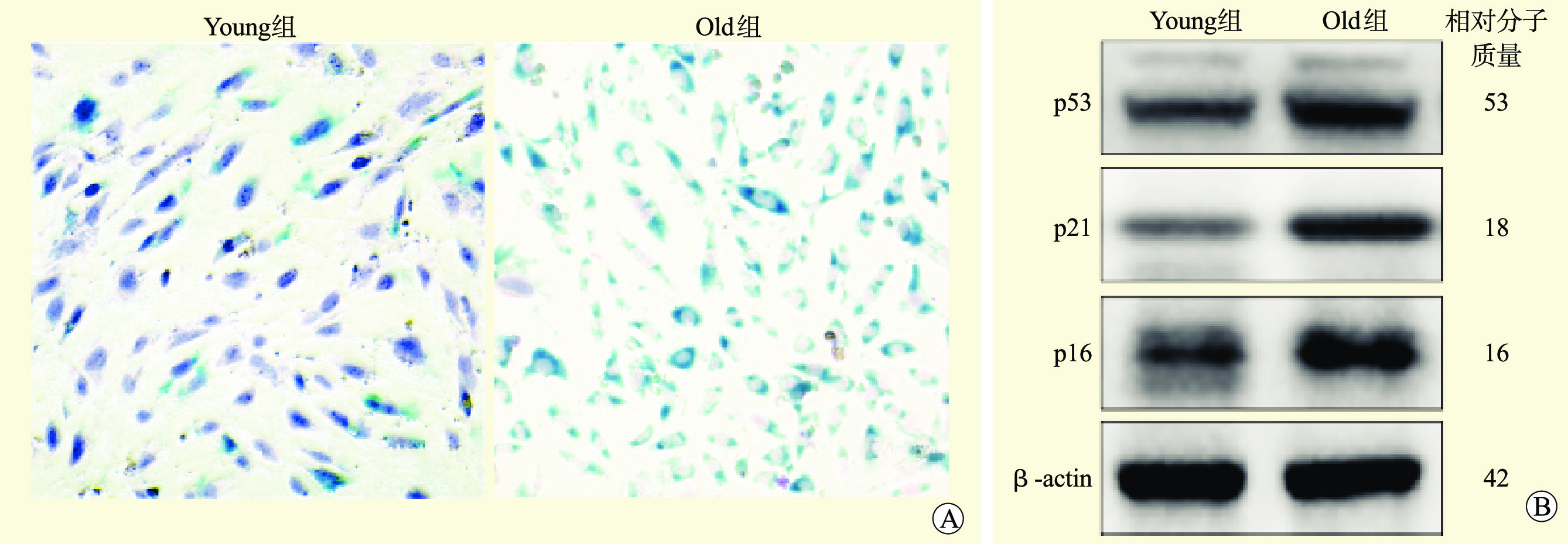
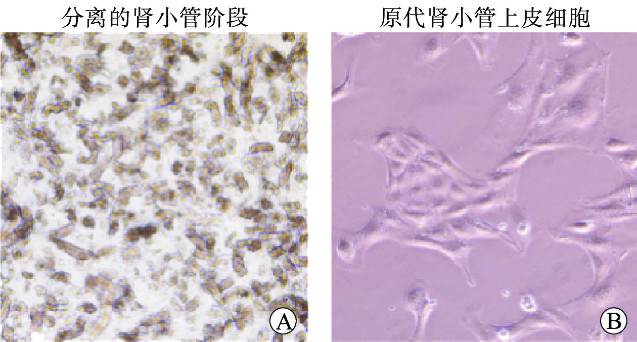
图 2 衰老细胞鉴定
注:A图为SA-β-gal染色(×100);B图为蛋白质印迹法。
Figure 2. Identification of senescent cell

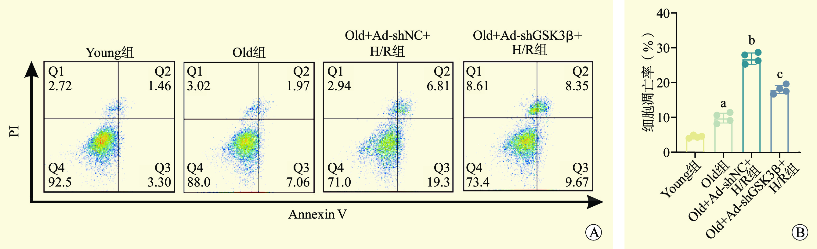
图 3 各组细胞凋亡水平
注:A图为流式细胞术检测各组细胞凋亡水平;B图为各组细胞凋亡率分析。与Young组比较,aP<0.05;与Old组比较,bP<0.05;与Old+Ad-shNC+H/R组比较,cP<0.05。
Figure 3. Apoptosis levels in each group

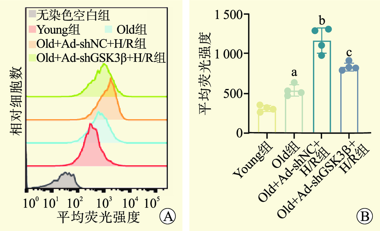
图 4 各组线粒体活性氧水平
注:A图为流式细胞术分析线粒体活性氧水平;B图为各组线粒体活性氧水平分析。与Young组比较,aP<0.05,与Old组比较,bP<0.05,与Old+Ad-shNC+H/R组比较,cP<0.05。
Figure 4. Mitochondrial reactive oxygen species levels in each group

图 6 各组GSK3β和MAM相关蛋白表达水平
Figure 6. The expression levels of GSK3β and MAM-associated proteins in each group
-
[1] YU S, LONG JJ, YU Y, et al. Survival benefit of accepting kidneys from older donation after cardiac death donors[J]. Am J Transplant, 2021, 21(3): 1138-1146. DOI: 10.1111/ajt.16198. [2] 邬莉萍, 张曙伟. 老年活体供肾移植现状与研究进展[J]. 现代实用医学, 2022, 34(11): 1403-1405. DOI: 10.3969/j.issn.1671-0800.2022.11.002.WU LP, ZHANG SG. Current status and research progress of elderly living donor kidney transplantation[J]. Mod Pract Med, 2022, 34(11): 1403-1405. DOI: 10.3969/j.issn.1671-0800.2022.11.002. [3] 潘佳善, 苏涌, 朱道方, 等. 公民逝世捐献与活体捐献肾移植的近期临床效果[J]. 实用医学杂志, 2022, 38(2): 184-189. DOI: 10.3969/j.issn.1006-5725.2022.02.011.PAN JS, SU Y, ZHU DF, et al. Clinical effects of deceased vs living donor on kidney transplantation[J]. J Pract Med, 2022, 38(2): 184-189. DOI: 10.3969/j.issn.1006-5725.2022.02.011. [4] SUMMERS DM, WATSON CJ, PETTIGREW GJ, et al. Kidney donation after circulatory death (DCD): state of the art[J]. Kidney Int, 2015, 88(2): 241-249. DOI: 10.1038/ki.2015.88. [5] PÉREZ-SÁEZ MJ, MONTERO N, REDONDO-PACHÓN D, et al. Strategies for an expanded use of kidneys from elderly donors[J]. Transplantation, 2017, 101(4): 727-745. DOI: 10.1097/TP.0000000000001635. [6] PRUETT TL, VECE GR, CARRICO RJ, et al. US deceased kidney transplantation: estimated GFR, donor age and KDPI association with graft survival[J]. EClinicalMedicine, 2021, 37: 100980. DOI: 10.1016/j.eclinm.2021.100980. [7] REX N, MELK A, SCHMITT R. Cellular senescence and kidney aging[J]. Clin Sci (Lond), 2023, 137(24): 1805-1821. DOI: 10.1042/CS20230140. [8] DONATE-CORREA J, MARTÍN-CARRO B, CANNATA-ANDÍA JB, et al. Klotho, oxidative stress, and mitochondrial damage in kidney disease[J]. Antioxidants (Basel), 2023, 12(2): 239. DOI: 10.3390/antiox12020239. [9] CHAUDHARY MR, CHAUDHARY S, SHARMA Y, et al. Aging, oxidative stress and degenerative diseases: mechanisms, complications and emerging therapeutic strategies[J]. Biogerontology, 2023, 24(5): 609-662. DOI: 10.1007/s10522-023-10050-1. [10] DENG LC, ALINEJAD T, BELLUSCI S, et al. Fibroblast growth factors in the management of acute kidney injury following ischemia-reperfusion[J]. Front Pharmacol, 2020, 11: 426. DOI: 10.3389/fphar.2020.00426. [11] TANG C, DONG Z. Mitochondria in kidney injury: when the power plant fails[J]. J Am Soc Nephrol, 2016, 27(7): 1869-1872. DOI: 10.1681/ASN.2015111277. [12] FUJII S, USHIODA R, NAGATA K. Redox states in the endoplasmic reticulum directly regulate the activity of calcium channel, inositol 1, 4, 5-trisphosphate receptors[J]. Proc Natl Acad Sci U S A, 2023, 120(22): e2216857120. DOI: 10.1073/pnas.2216857120. [13] YUAN M, GONG M, HE J, et al. IP3R1/GRP75/VDAC1 complex mediates endoplasmic reticulum stress-mitochondrial oxidative stress in diabetic atrial remodeling[J]. Redox Biol, 2022, 52: 102289. DOI: 10.1016/j.redox.2022.102289. [14] ZIEGLER DV, VINDRIEUX D, GOEHRIG D, et al. Calcium channel ITPR2 and mitochondria-ER contacts promote cellular senescence and aging[J]. Nat Commun, 2021, 12(1): 720. DOI: 10.1038/s41467-021-20993-z. [15] MORCIANO G, GIORGI C, BONORA M, et al. Molecular identity of the mitochondrial permeability transition pore and its role in ischemia-reperfusion injury[J]. J Mol Cell Cardiol, 2015, 78: 142-153. DOI: 10.1016/j.yjmcc.2014.08.015. [16] FANG Y, CHEN B, LIU Z, et al. Age-related GSK3β overexpression drives podocyte senescence and glomerular aging[J]. J Clin Invest, 2022, 132(4): e141848. DOI: 10.1172/JCI141848. [17] KUANG BC, WANG ZH, HOU SH, et al. Methyl eugenol protects the kidney from oxidative damage in mice by blocking the Nrf2 nuclear export signal through activation of the AMPK/GSK3β axis[J]. Acta Pharmacol Sin, 2023, 44(2): 367-380. DOI: 10.1038/s41401-022-00942-2. [18] COELLO I, MARTÍNEZ AI, PERAIRE M, et al. Effect of ischemia times and donor and recipient features on Maastricht category III kidney transplant outcomes[J]. Arch Esp Urol, 2022, 75(7): 612-617. DOI: 10.56434/j.arch.esp.urol.20227507.88. [19] LIM WH, OOI E, PILMORE HL, et al. Interactions between donor age and 12-month estimated glomerular filtration rate on allograft and patient outcomes after kidney transplantation[J]. Transpl Int, 2022, 35: 10199. DOI: 10.3389/ti.2022.10199. [20] SCHMITT R, MELK A. Molecular mechanisms of renal aging[J]. Kidney Int, 2017, 92(3): 569-579. DOI: 10.1016/j.kint.2017.02.036. [21] MAROSI M, ARMAN P, ACETO G, et al. Glycogen synthase kinase 3: ion channels, plasticity, and diseases[J]. Int J Mol Sci, 2022, 23(8): 4413. DOI: 10.3390/ijms23084413. [22] 许艳玲, 赵玉珠, 付裕, 等. 青蒿琥酯通过PI3K/GSK-3β通路对1型糖尿病小鼠胰岛素抵抗的改善作用研究[J]. 天津中医药, 2022, 39(8): 1077-1081. DOI: 10.11656/j.issn.1672-1519.2022.08.23.XU YL, ZHAO YZ, FU Y, et al. Effect of artesunate on insulin resistance in type 1 diabetic mouse through PI3K/GSK-3β pathway[J]. Tianjin J Tradit Chin Med, 2022, 39(8): 1077-1081. DOI: 10.11656/j.issn.1672-1519.2022.08.23. [23] KREIDBERG JA, SCHUMACHER VA. GSK3β and the aging kidney[J]. J Clin Invest, 2022, 132(4): e155885. DOI: 10.1172/JCI155885. [24] 信强, 崔碧红, 苏秀兰, 等. 缺血再灌注对老年心脏的损伤机制及研究进展[J]. 内蒙古医科大学学报, 2023, 45(1): 101-105.XIN Q, CUI BH, SU XL, et al. Mechanism and research progress of ischemia-reperfusion injury in elderly heart[J]. J Inner Mongolia Med Univ, 2023, 45(1): 101-105. [25] CHEN Q, SONG Y, YANG N, et al. Aging deteriorated liver ischemia and reperfusion injury by suppressing Tribble's proteins 1 mediated macrophage polarization[J]. Bioengineered, 2022, 13(6): 14519-14533. DOI: 10.1080/21655979.2022.2090218. [26] KOSTYAK JC, HUNTER JC, KORZICK DH. Acute PKCdelta inhibition limits ischaemia-reperfusion injury in the aged rat heart: role of GSK-3beta[J]. Cardiovasc Res, 2006, 70(2): 325-334. DOI: 10.1016/j.cardiores.2006.02.009. [27] KORZICK DH, KOSTYAK JC, HUNTER JC, et al Local delivery of PKCepsilon-activating peptide mimics ischemic preconditioning in aged hearts through GSK-3beta but not F1-ATPase inactivation[J]. Am J Physiol Heart Circ Physiol, 2007, 293(4): H2056-H2063. DOI: 10.1152/ajpheart.00403.2007. [28] NELSON PJ, CANTLEY L. GSK3beta plays dirty in acute kidney injury[J]. J Am Soc Nephrol, 2010, 21(2): 199-200. DOI: 10.1681/ASN.2009121214. [29] ZHOU S, WANG P, QIAO Y, et al. Genetic and pharmacologic targeting of glycogen synthase kinase 3β reinforces the Nrf2 antioxidant defense against podocytopathy[J]. J Am Soc Nephrol, 2016, 27(8): 2289-2308. DOI: 10.1681/ASN.2015050565. [30] SUN Y, FAN Y, WANG Z, et al. S100A16 promotes acute kidney injury by activating HRD1-induced ubiquitination and degradation of GSK3β and CK1α[J]. Cell Mol Life Sci, 2022, 79(3): 184. DOI: 10.1007/s00018-022-04213-5. [31] GUO J, ZHENG W, LIU Y, et al. Long non-coding RNA DLX6-AS1 is the key mediator of glomerular podocyte injury and albuminuria in diabetic nephropathy by targeting the miR-346/GSK-3β signaling pathway[J]. Cell Death Dis, 2023, 14(2): 172. DOI: 10.1038/s41419-023-05695-2. [32] CHEN B, WANG P, LIANG X, et al. Permissive effect of GSK3β on profibrogenic plasticity of renal tubular cells in progressive chronic kidney disease[J]. Cell Death Dis, 2021, 12(5): 432. DOI: 10.1038/s41419-021-03709-5. [33] ZENG L, NG JK, FUNG WW, et al. Intrarenal and urinary glycogen synthase kinase-3 beta levels in diabetic and nondiabetic chronic kidney disease[J]. Kidney Blood Press Res, 2023, 48(1): 241-248. DOI: 10.1159/000530210. [34] JANIKIEWICZ J, SZYMAŃSKI J, MALINSKA D, et al. Mitochondria-associated membranes in aging and senescence: structure, function, and dynamics[J]. Cell Death Dis, 2018, 9(3): 332. DOI: 10.1038/s41419-017-0105-5. [35] FUJIMOTO T, SHIRASAWA S. Identification of KRAP-expressing cells and the functional relevance of KRAP to the subcellular localization of IP3R in the stomach and kidney[J]. Int J Mol Med, 2012, 30(6): 1287-1293. DOI: 10.3892/ijmm.2012.1126. [36] LIU Y, MA X, FUJIOKA H, et al. DJ-1 regulates the integrity and function of ER-mitochondria association through interaction with IP3R3-GRP75-VDAC1[J]. Proc Natl Acad Sci U S A, 2019, 116(50): 25322-25328. DOI: 10.1073/pnas.1906565116. [37] GOMEZ L, THIEBAUT PA, PAILLARD M, et al. The SR/ER-mitochondria calcium crosstalk is regulated by GSK3β during reperfusion injury[J]. Cell Death Differ, 2016, 23(2): 313-322. DOI: 10.1038/cdd.2015.101. [38] PASTORINO JG, HOEK JB, SHULGA N. Activation of glycogen synthase kinase 3beta disrupts the binding of hexokinase II to mitochondria by phosphorylating voltage-dependent anion channel and potentiates chemotherapy-induced cytotoxicity[J]. Cancer Res, 2005, 65(22): 10545-10554. DOI: 10.1158/0008-5472.CAN-05-1925. [39] DAS S, WONG R, RAJAPAKSE N, et al. Glycogen synthase kinase 3 inhibition slows mitochondrial adenine nucleotide transport and regulates voltage-dependent anion channel phosphorylation[J]. Circ Res, 2008, 103(9): 983-991. DOI: 10.1161/CIRCRESAHA.108.178970. [40] THOUDAM T, CHANDA D, LEE JY, et al. Enhanced Ca2+-channeling complex formation at the ER-mitochondria interface underlies the pathogenesis of alcohol-associated liver disease[J]. Nat Commun, 2023, 14(1): 1703. DOI: 10.1038/s41467-023-37214-4. -

 下载:
下载:


 点击查看大图
计量
- 文章访问数: 21
- HTML全文浏览量: 14
- PDF下载量: 4
- 被引次数: 0