Clinical analysis of 18 patients with portal vein stenosis after liver transplantation
-
摘要:
目的 总结肝移植术后门静脉狭窄的诊治经验。 方法 回顾性分析18例肝移植术后门静脉狭窄患者的临床资料,总结门静脉狭窄发生情况、治疗情况及预后。 结果 17例患者肝移植术前有肝硬化病史,7例患者肝移植术前有门静脉血栓形成史,8例有脾切除断流或分流等相关手术史;3例为儿童供肝。18例患者门静脉狭窄发生时间为术后23 d~24个月,中位时间为2.2个月,均经彩色多普勒超声(彩超)检查发现,通过门静脉CT血管造影术(CTA)或行介入治疗确诊。所有病例确诊后,均行华法林抗凝治疗;5例有门静脉高压表现的患者行球囊扩张术,其中1例同时放置血管内支架;其余13例采取保守治疗。治疗后9例好转,7例无变化,2例加重。 结论 对肝移植术前有肝硬化病史的受者,术后常规彩超监测,CTA或介入治疗确诊门静脉狭窄。无临床症状患者可行保守治疗,合并门静脉高压者可行介入治疗,多数病例预后良好。 Abstract:Objective To summarize the experience of clinical diagnosis and treatment of portal vein stenosis after liver transplantation. Methods Clinical data of 18 patients presenting with portal vein stenosis after undergoing liver transplantation were retrospectively analyzed. The incidence, treatment and prognosis of portal vein stenosis were summarized. Results Seventeen patients had a medical history of liver cirrhosis before liver transplantation, 7 cases with a medical history of portal vein thrombosis and 8 cases with a medical history of devascularization or shunt with splenectomy. Three cases received the pediatric liver grafts. Eighteen patients suffered from portal vein stenosis from postoperative 23 d to 24 months with a median time of 2.2 months, which was detected by color Doppler ultrasound (CDU) and diagnosed by CT angiography (CTA) of the portal vein or interventional therapy. After the diagnosis was confirmed, all cases received anticoagulant treatment by warfarin. Five patients with portal hypertension underwent balloon dilatation, and one of them received endovascular stent implantation simultaneously. The remaining 13 patients received conservative therapy. After corresponding treatment, 9 cases were mitigated, 7 patients remained unchanged and 2 cases were aggravated. Conclusions For the recipients with a medical history of liver cirrhosis before liver transplantation, portal vein stenosis should be monitored by conventional CDU and diagnosed by CTA or interventional therapy after transplantation. Patients without clinical symptoms can receive conservative treatment. Those complicated with portal hypertension can undergo interventional therapy. Favorable clinical prognosis is obtained in most cases. -

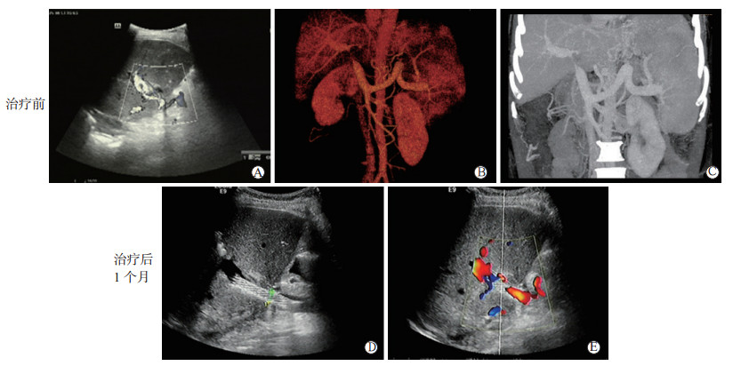
图 1 1例门静脉狭窄患者治疗前后的影像学表现
A图为治疗前彩超检查示门静脉主干内径10 mm,为双相血流,入肝瞬时流速209 cm/s,出肝瞬时流速40 cm/s,门静脉吻合口内径2.7 mm,瞬时流速146 cm/s;B图和C图为治疗前CTA显示第一肝门部门静脉主干被稍高密度影包绕,致局部管腔重度狭窄,门静脉高压,胃底静脉增粗迂曲,汇入脾静脉,胃左静脉可见分支与左肾静脉相沟通,门静脉左支主干及属支管腔内低密度充盈缺损影,考虑附壁血栓形成;D图为治疗后1个月彩超显示门静脉主干内可见支架回声,支架外径约8.4 mm,支架内暗区清晰;E图为治疗后1个月彩超显示支架血流充盈良好,瞬时流速46 cm/s
Figure 1. Imaging findings of one patient with portal vein stenosis before and after treatment
-
[1] Huang TL, Cheng YF, Chen TY, et al. Doppler ultrasound evaluation of postoperative portal vein stenosis in adult living donor liver transplantation[J]. Transplant Proc, 2010, 42(3) : 879-881. DOI: 10.1016/j.transproceed.2010.02.036. [2] Funaki B, Rosenblum JD, Leef JA, et al. Percutaneous treatment of portal venous stenosis in children and adolescents with segmental hepatic transplants:long-term results[J]. Radiology, 2000, 215(1): 147-151.DOI: 10.1148/radiology.215.1.r00ap38147. [3] Chang WT, Kuo YT, Lee KT, et al. The value of primary vascular stents in management of early portal vein stenosis after liver transplantation[J]. Kaohsiung J Med Sci, 2016, 32(3): 128-134. DOI: 10.1016/j.kjms.2016.02.003. [4] 何晓顺, 鞠卫强, 赵强.公民器官捐献时代肝脏移植常见并发症的处理[J].中国普外基础与临床杂志, 2015, 22(12): 1417-1419. doi: 10.7507/1007-9424.20150371He XS, Ju WQ, Zhao Q. The treatment of common complications of liver transplantation in the age of donation after citizen's death[J]. Chin J Bases Clin Gen Surg, 2015, 22(12): 1417-1419. doi: 10.7507/1007-9424.20150371 [5] 杨翰, 张水军. 肝移植术后血管并发症及其治疗进展[J/CD]. 实用器官移植电子杂志, 2014, 2(5): 317-320. DOI: 10.3969/j.issn.2095-5332.2014.05.012.Yang H, Zhang SJ. The treatment progress and vascular complications after liver transplantation[J/CD]. Pract J Organ Transplant (Electr Vers), 2014, 2(5): 317-320. DOI: 10.3969/j.issn.2095-5332.2014.05.012. [6] Ma L, Lu Q, Luo Y. Vascular complications after adult living donor liver transplantation: evaluation with ultrasonography[J]. World J Gastroenteral, 2016, 22(4): 1617-1626. DOI: 10.3748/wjg.v22.i4.1617. [7] 王晓静, 王岩青, 郭朝锋, 等.肝移植术后门静脉并发症的超声检测价值[J].临床超声医学杂志, 2017, 19(5): 341-343. DOI: 10.3969/j.issn.1008-6978.2017.05.022.Wang XJ, Wang YQ, Guo CF, et al. Clinical value of color Doppler ultrasound in detection of portal vein complications after liver transplantation[J]. J Clin Ultrasound Med, 2017, 19(5): 341-343. DOI: 10.3969/j.issn.1008-6978.2017.05.022. [8] 汪明月, 鲁植艳.肝移植术后并发症的影像学评估[J].临床肝胆病杂志, 2016, 32(12): 2295-2299. DOI: 10.3969/j.issn.1001-5256.2016.12.013.Wang MY, Lu ZY. Imaging evaluation of complications after liver transplantation[J]. J Clin Hepatol, 2016, 32(12): 2295-2299. DOI: 10.3969/j.issn.1001-5256.2016.12.013. [9] Olcott EW, Ring EJ, Roberts JP, et al. Percutaneous transhepatic portal vein angioplasty and stent placement after liver transplantation: early experience[J]. J Vasc Interv Radiol, 1990, 1(1): 17-22. DOI: 10.1016/S1051-0443(90)72496-7. [10] Wang JF, Zhai RY, Wei BJ, et al. Percutaneous intravascular stents for treatment of portal venous stenosis after liver transplantation: midterm results[J]. Transplant Proc, 2006, 38(5): 1461-1462.DOI: 10.1016/j.transproceed.2006.02.113. [11] Uller W, Knoppke B, Schreyer AG, et al. Interventional radiological treatment of perihepatic vascular stenosis or occlusion in pediatric patients after liver transplantation[J]. Cardiovasc Interv Radiol, 2013, 36(6): 1562-1571. DOI: 10.1007/s00270-013-0595-1. [12] Ohm JY, Ko GY, Sung KB, et al. Safety and efficacy of transhepatic and transsplenic access for endovascular management of portal vein complications after liver transplantation[J]. Liver Transpl, 2017, 23(9): 1133-1142. DOI: 10.1002/lt.24737. [13] Yabuta M, Shibata T, Shibata T, et al. Long-term outcome of percutaneous transhepatic balloon angioplasty for portal vein stenosis after pediatric living donor liver transplantation: a single institute's experience[J]. J Vasc Interv Radiol, 2014, 25(9): 1406-1412. DOI: 10.1016/j.jvir.2014.03.034. [14] Gao H, Wang H, Chen G, et al. Intervention therapy for portal vein stenosis /occlusion after pediatric liver transplantation[J]. Ann Transplant, 2017, 18(22): 222-229. DOI: 10.12659/AOT.902239. [15] 刘煜, 牛玉坚, 任秀昀, 等.肝移植后门静脉供血障碍的诊断和治疗[J].中华器官移植杂志, 2006, 27(10): 619-620. DOI: 10.3760/cma.j.issn.0254-1785.2006.10.012.Liu Y, Niu YJ, Ren XY, et al. Diagnosis and treatment for the insufficient supply to the portal vein after liver transplantation[J]. Chin J Organ Transplant, 2006, 27(10): 619-620. DOI: 10.3760/cma.j.issn.0254-1785.2006.10.012. [16] 孙军辉, 周官辉, 郑树森.肝移植介入治疗应用及进展[J].器官移植, 2015, 6(3): 134-138. DOI: 10.3969/j.issn.1674-7445.2015.03.002.Sun JH, Zhou GH, Zheng SS. Application and progress of interventional therapy in liver transplantation[J]. Organ Transplant, 2015, 6(3): 134-138. DOI: 10.3969/j.issn.1674-7445.2015.03.002. [17] Mullan CP, Siewert B, Kane RA, et al. Can Doppler sonography discern between hemodynamically significant and insignificant portal vein stenosis after adult liver transplantation?[J]. Am J Roentgenol, 2010, 195(6):1438-1443. DOI: 10.2214/AJR.10.4636. [18] Wang JF, Yang WL, Huang Q, et al. Interventional treatment for portal venous occlusion after liver transplantation: long-term follow-up results[J]. Medicine, 2015, 94(4): e356. DOI: 10.1097/MD.0000000000000356. -

 下载:
下载:

 点击查看大图
图(1)
计量
- 文章访问数: 152
- HTML全文浏览量: 94
- PDF下载量: 14
- 被引次数: 0