Determination of optimal threshold for hepatic artery stenosis on Doppler ultrasonography and its effect for clinical decision of treatment for patients with tardus parvus waveform after liver transplantation
-
摘要:
目的 分析多普勒超声(DUS)诊断肝移植术后肝动脉狭窄(HAS)的最佳临界值,并结合肝功能异常,提出小慢波(TPW)患者接受CT血管造影(CTA)或数字血管造影(DSA)检查的诊断标准。 方法 收集171例行肝移植术并在术后常规复查DUS、肝功能检查并行CTA或DSA检查确诊患者的临床资料。采用多水平似然比(MLR)确定肝动脉阻力指数(RI)及收缩期加速时间(SAT)诊断HAS的最佳临界值。建立不同诊断标准(低信心类为TPW阳性,中等信心类为TPW阳性+肝功能异常;高信心类为TPW阳性+肝功能异常或TPW阳性+最佳临界值)并比较其诊断效能。 结果 MLR显示诊断HAS的最佳临界值为RI < 0.4,SAT>0.12 s。中等信心类及高信心类诊断标准的特异度明显高于低信心类(P < 0.05),且假阳性率明显降低(P < 0.05)。另外,中等信心类诊断标准的灵敏度明显低于低信心类及高信心类(P < 0.05),而低信心类与高信心类诊断标准间的灵敏度差异无统计学意义(P>0.05)。 结论 对于肝移植术后DUS检查显示TPW阳性的患者,结合肝功能异常及最佳临界值这一诊断标准可帮助临床医师对其作出适当的临床决策。 Abstract:Objective To analyze the optimal threshold of Doppler ultrasonography (DUS) in the diagnosis of hepatic artery stenosis (HAS) after liver transplantation and propose the diagnostic criteria of CT angiography (CTA) or digital subtraction angiography (DSA) for patients with tardus parvus waveform (TPW) in combination with liver dysfunction. Methods Clinical data of 171 patients undergoing liver transplantation, postoperative conventional DUS, liver function test, CTA or DSA were collected. The optimal threshold of resistance index (RI) and systolic acceleration time (SAT) for the diagnosis of HAS were determined by multi-level likelihood ratio (MLR). Different diagnostic criteria were established and the diagnostic efficacy was statistically compared. Positive TPW was defined as the diagnostic criterion with low confidence, positive TPW + liver dysfunction as the moderate confidences, and positive TPW + liver dysfunction or positive TPW + optimal threshold as the high confidence. Results MLR revealed that RI < 0.4 and SAT>0.12 s were the optimal threshold for the diagnosis of HAS. The specificity of diagnostic criteria with moderate and high confidence was significantly higher compared with that of the low confidence (P < 0.05). Moreover, the false-positive rate was significantly decreased (P < 0.05). The sensitivity of diagnostic criterion with moderate confidence was significantly lower than those of low and high confidence (both P < 0.05), whereas the sensitivity did not significantly differ between the diagnostic criteria with low and high confidence (P>0.05). Conclusions For patients with positive TPW detected by DUS after liver transplantation, the optimal threshold of diagnostic criteria combined with liver dysfunction contribute to appropriate clinical decision-making for clinicians. -

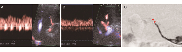
图 1 1例肝移植术后肝动脉狭窄患者的超声图像和DSA显影情况
本例患者,男性,26岁。肝衰竭行原位肝移植术后约4个月复查,多普勒超声显示肝左动脉RI 0.28,SAT 0.11 s(A图),肝右动脉RI 0.31,SAT 0.11s(B图),肝功能各项指标正常,符合高信心类诊断标准,最后经DSA证实肝动脉吻合口重度狭窄(C图,红色箭头所示),肝内动脉显影浅淡
Figure 1. Doppler ultrasonography and DSA of a case with hepatic artery stenosis after liver transplantation
表 1 肝动脉狭窄及非肝动脉狭窄患者一般情况及DUS检查结果的比较
Table 1. Comparison of the general situation and DUS examination results of patients with hepatic artery stenosis and non hepatic artery stenosis
指标 HAS组
(n=67)非HAS组
(n=105)P值 年龄(岁) 46±10 47±13 0.07 男性[n (%)] 59 (88) 88 (83.8) 0.51 小慢波[n (%)] 42 (63) 7(7.7) < 0.01 最小RI 0.45±0.13 0.62±0.10 < 0.01 最大SAT (s) 0.11±0.04 0.07±0.02 < 0.01  下载: 导出CSV
表 2 多水平似然比分析多普勒超声诊断肝动脉狭窄最佳临界值
Table 2. Multi level likelihood ratio analysis on optimal critical value of Doppler ultrasonography in the diagnosis of hepatic artery stenosis
指标 LR值 最小RI < 0.40 10.58 0.40~0.50 3.52 0.51~0.70 0.83 > 0.70 0.24 最大SAT (s) < 0.06 0.19 0.06~0.08 0.66 0.09~0.12 3.70 > 0.12 16.46
下载: 导出CSV
表 3 不同诊断标准的效能比较
Table 3. Comparison of efficiency in different diagnosis categories
标准 灵敏度 特异度 准确度 假阳性率(%) 低信心类 0.746 0.819 0.791 18.1 中等信心类 0.418 0.924 0.727 7.6 高信心类 0.612 0.981 0.837 1.9
下载: 导出CSV
-
[1] Gad EH, Abdelsamee MA, Kamel Y. Hepatic arterial and portal venous complications after adult and pediatric living donor liver transplantation, risk factors, management and outcome (a retrospective cohort study) [J]. Ann Med Surg, 2016, 8: 28-39. DOI: 10.1016/j.amsu.2016.04.021. [2] Pakosz-Golanowsha M, Lubikowski J, Post M, et al. The arterial anastomosis in liver transplantation: complications, treatment and outcome [J]. Hepatogastroenterology, 2010, 57(104): 1477-1482. https://www.researchgate.net/publication/50890242_The_Arterial_Anastomosis_in_Liver_Transplantation_Complications_Treatment_and_Outcome [3] Sanada Y, Wakiya T, Hishikawa S, et al. Risk factors and treatments for hepatic arterial complications in pediatric living donor liver transplantation [J]. J Hepatobiliary Pancreat Sci, 2014, 21(7): 463-472. DOI: 10.1002/jhbp.49. [4] Hamby BA, Ramirez DE, Loss GE, et al. Endovascular treatment of hepatic artery stenosis after liver transplantation [J]. J Vasc Surg, 2013, 57(4): 1067-1072. DOI: 10.1016/j.jvs.2012.10.086. [5] Le L, Terral W, Zea N, et al. Primary stent placement for hepatic artery stenosis after liver transplantation [J]. J Vasc Surg, 2015, 62(3): 704-709. DOI: 10.1016/j.jvs.2015.04.400. [6] Nemes B, Gaman G, Sárváry E, et al. Retransplantations in the Hungarian liver transplant program [J]. Transplant Proc, 2013, 45(10): 3688-3690. DOI: 10.1016/j.transproceed.2013.10.004. [7] Rostambeigi N, Hunter D, Duval S, et al. Stent placement versus angioplasty for hepatic artery stenosis after liver transplant: a meta-analysis of case series [J]. Eur Radiol, 2013, 23(5): 1323-1334. DOI: 10.1007/s00330-012-2730-9. [8] Uller W, Knoppke B, Schreyer AG, et al. Interventional radiological treatment of perihepatic vascular stenosis or occlusion in pediatric patients after liver transplantation [J]. Cardiovasc Intervent Radiol, 2013, 36(6): 1562-1571. DOI: 10.1007/s00270-013-0595-1. [9] Vidjak V, Novačić K, Matijević F, et al. Percutaneous endovascular treatment for hepatic artery stenosis after liver transplantation: the role of percutaneous endovascular treatment [J]. Pol J Radiol, 2015, 80: 309-316. DOI: 10.12659/PJR.893831. [10] Dodd GD 3rd, Memel DS, Zajko AB, et al. Hepatic artery stenosis and thrombosis in transplant recipients: Doppler diagnosis with resistive index and systolic acceleration time [J]. Radiology, 1994, 192(3): 657-661. doi: 10.1148/radiology.192.3.8058930 [11] Park YS, Kim KW, Lee SJ, et al. Hepatic arterial stenosis assessed with doppler US after liver transplantation: frequent false-positive diagnoses with tardus parvus waveform and value of adding optimal peak systolic velocity cutoff [J]. Radiology, 2011, 260(3): 884-891. DOI: 10.1148/radiol.11102257. [12] Deeks JJ, Altman DG. Diagnostic tests 4: likelihood ratios [J]. BMJ, 2004, 329(7458): 168-169. doi: 10.1136/bmj.329.7458.168 [13] Grimes DA, Schulz KF. Refining clinical diagnosis with likelihood ratios [J]. Lancet, 2005, 365(9469): 1500-1505. doi: 10.1016/S0140-6736(05)66422-7 [14] Platt JF, Yutzy GG, Bude RO, et al. Use of Doppler sonography for revealing hepatic artery stenosis in liver transplant recipients[J]. AJR Am J Roentgenol, 1997, 168(2): 473-476. doi: 10.2214/ajr.168.2.9016229 [15] Sidhu PS, Ellis SM, Karani JB, et al. Hepatic artery stenosis following liver transplantation: significance of the tardus parvus waveform and the role of microbubble contrast media in the detection of a focal stenosis [J]. Clin Radiol, 2002, 57(9): 789-799. doi: 10.1016/S0009-9260(02)90969-4 [16] Vit A, De Candia A, Como G, et al. Doppler evaluation of arterial complications of adult orthotopic liver transplantation[J]. J Clin Ultrasound, 2003, 31(7): 339-345. doi: 10.1002/(ISSN)1097-0096 [17] De Gaetano AM, Cotroneo AR, Maresca G, et al. Color Doppler sonography in the diagnosis and monitoring of arterial complications after liver transplantation [J]. J Clin Ultrasound, 2000, 28(8): 373-380. doi: 10.1002/(ISSN)1097-0096 [18] Maceneaney PM, Malone DE, Skehan SJ, et al. The role of hepatic arterial Doppler ultrasound after liver transplantation: an 'audit cycle' evaluation [J]. Clin Radiol, 2000, 55(7): 517-524. doi: 10.1053/crad.1999.0486 [19] Tamsel S, Demirpolat G, Killi R, et al. Vascular complications after liver transplantation: evaluation with Doppler US [J]. Abdominal Imaging, 2007, 32(3): 339-347. doi: 10.1007/s00261-006-9041-z [20] Garcia-Criado A, Gilabert R, Berzigotti A, et al. Doppler ultrasound findings in the hepatic artery shortly after liver transplantation [J]. AJR Am J Roentgenol, 2009, 193(1): 128-135. DOI: 10.2214/AJR.07.3919. [21] Choi EK, Lu DS, Park SH, et al. Doppler US for suspicion of hepatic arterial ischemia in orthotopically transplanted livers: role of central versus intrahepatic waveform analysis [J]. Radiology, 2013, 267(1): 276-284. DOI: 10.1148/radiol.12120557. -

 下载:
下载:

 点击查看大图
计量
- 文章访问数: 150
- HTML全文浏览量: 64
- PDF下载量: 7
- 被引次数: 0