Mechanism of human umbilical cord mesenchymal stem cells alleviating ischemia-reperfusion injury of hepatocytes through mitochondrial transfer
-
摘要:
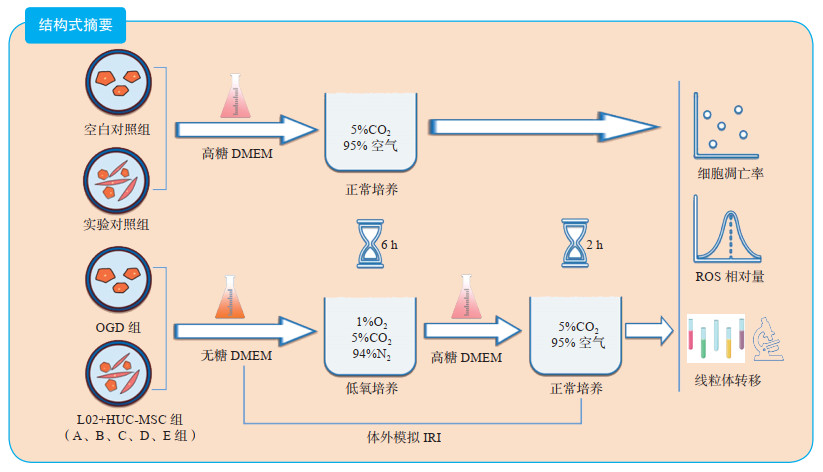
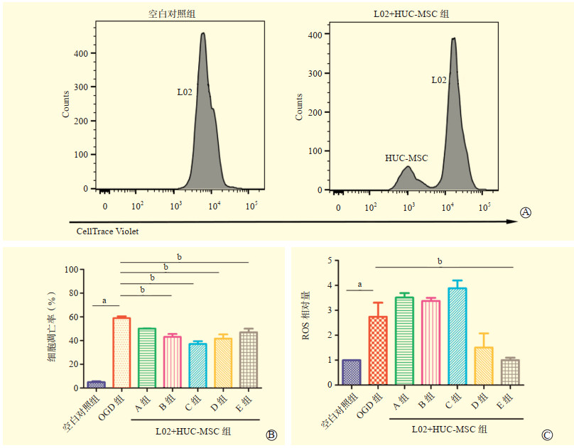
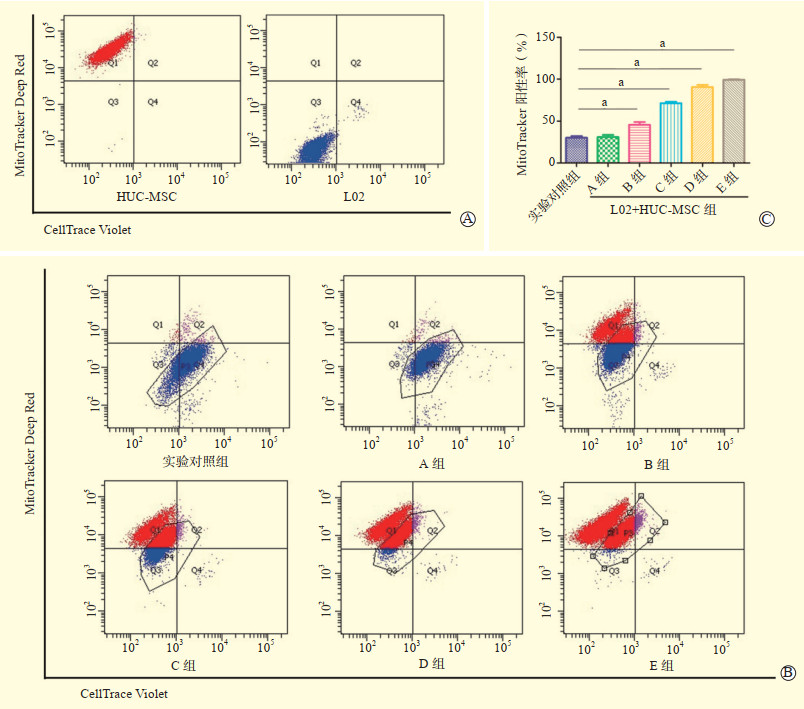
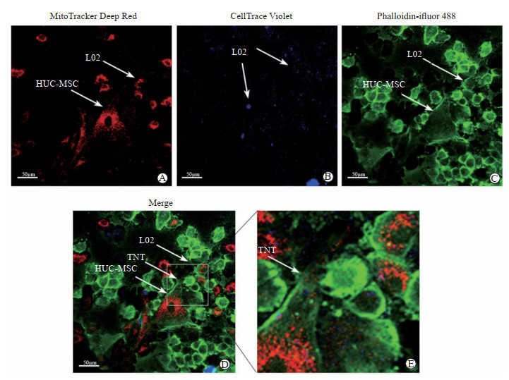
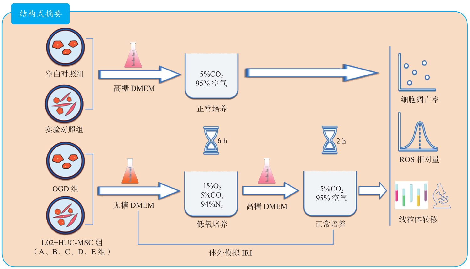
目的 探讨人脐带间充质干细胞(HUC-MSC)通过线粒体转移减轻肝细胞缺血-再灌注损伤(IRI)的作用机制。 方法 将正常人肝细胞株L02分为空白对照组、氧糖剥夺(OGD)组、实验对照组、L02与HUC-MSC共培养组(L02+HUC-MSC组),其中L02+HUC-MSC组根据L02与HUC-MSC不同共培养比例分为10:1共培养组(A组)、4:1共培养组(B组)、2:1共培养组(C组)、1:1共培养组(D组)和1:2共培养组(E组)。采用流式细胞术检测L02细胞凋亡率和细胞内活性氧簇(ROS)相对量;采用流式细胞术检测L02的MitoTracker阳性率;采用激光共聚焦显微镜观察HUC-MSC向L02转移线粒体的情况。 结果 OGD组L02细胞凋亡率和细胞内ROS相对量高于空白对照组(均为P < 0.05);与OGD组比较,B、C、D、E组L02细胞凋亡率均降低(均为P < 0.05),E组L02细胞内ROS相对量降低(P < 0.05)。A组L02的MitoTracker阳性率与实验对照组比较,差异无统计学意义(P>0.05),B、C、D、E组均较实验对照组增加(均为P < 0.05),并呈浓度依赖性。在激光共聚焦显微镜下可观察到线粒体通过隧道纳米管(TNT)从HUC-MSC向L02转移。 结论 HUC-MSC可通过细胞间直接转移线粒体的方式减轻肝细胞IRI后细胞凋亡和降低细胞内ROS水平。 -
关键词:
- 缺血-再灌注损伤(IRI) /
- 人脐带间充质干细胞(HUC-MSC) /
- 氧糖剥夺(OGD) /
- 活性氧簇(ROS) /
- 线粒体转移 /
- 隧道纳米管(TNT) /
- 细胞凋亡 /
- 免疫治疗
Abstract:Objective To explore the mechanism of human umbilical cord mesenchymal stem cell (HUC-MSC) alleviating ischemia-reperfusion injury (IRI) of liver cells through mitochondrial transfer. Methods Normal human liver cell line L02 was divided into the blank control group, oxygen-glucose deprivation (OGD) group, experimental control group, and L02 and HUC-MSC co-culture group (L02+HUC-MSC group). L02+HUC-MSC group was further divided into 10:1 co-culture subgroup (group A), 4:1 co-culture subgroup (group B), 2:1 co-culture subgroup (group C), 1:1co-culture subgroup (group D) and 1:2 co-culture subgroup (group E) according to different co-culture ratio of L02 and HUC-MSC. The apoptosis rate and relative reactive oxygen species (ROS) level of L02 cells were detected by flow cytometry. The MitoTracker positive rate of L02 cells was detected by flow cytometry. The mitochondrial transfer from HUC-MSC to L02 cells was observed by laser confocal microscope. Results The apoptosis rate and relative ROS level of L02 cells in the OGD group were significantly higher than those in the blank control group (both P < 0.05). Compared with the OGD group, the apoptosis rates of L02 cells in group B, C, D and E were significantly decreased (all P < 0.05), and the relative ROS level of L02 cells in group E was significantly declined (P < 0.05). The MitoTracker positive rate of L02 cells did not significantly differ between group A and experimental control group (P>0.05), whereas the MitoTracker positive rates of L02 cells in group B, C, D and E were significantly higher than that in the experimental control group in a concentration-dependent manner (all P < 0.05). Under the laser confocal microscope, mitochondrial transfer fromHUC-MSC to L02 cells could be observed through tunneling nanotube (TNT). Conclusions HUC-MSC may alleviate cell apoptosis and reduce ROS level of liver cells after IRI via direct mitochondrial transfer between cells. -

图 1 各组L02细胞凋亡率和细胞内ROS相对量的比较
注:A图为空白对照组和L02+HUC-MSC组的流式细胞图;B图为各组L02细胞凋亡率的比较;C图为各组L02细胞内ROS相对量的比较;与空白对照组比较,aP < 0.05,与OGD组比较,bP < 0.05。
Figure 1. Comparison of cell apoptosis rate and intracellular relative ROS level of L02 cells among each group

图 2 HUC-MSC向L02转移线粒体的流式细胞学表现
注:A图为流式细胞术检测HUC-MSC与L02;B图为各组HUC-MSC向L02转移线粒体的情况,圈门表示L02细胞群;C图为通过L02细胞群中MitoTracker阳性L02的比例来评估线粒体转移率,与实验对照组比较,aP < 0.05。
Figure 2. Flow cytometry findings of mitochondrial transfer from HUC-MSC to L02 cells

图 3 HUC-MSC向L02转移线粒体的激光共聚焦显微镜下表现
注:A图示MitoTracker Deep Red标记的HUC-MSC(×200);B图示CellTrace Violet标记的L02(×200);C图示Phaliodin-ifluor 488染色的F-actin(×200);D图(×200)、E图(×400)示HUC-MSC和L02共培养后TNT形成和线粒体转移。
Figure 3. Mitochondrial transfer from HUC-MSC to L02 cells under the confocal laser scanning microscopy
-
[1] HEYLEN L, PIRENNE J, NAESENS M, et al. "Time is tissue"-a minireview on the importance of donor nephrectomy, donor hepatectomy, and implantation times in kidney and liver transplantation[J]. Am J Transplant, 2021, DOI: 10.1111/ajt.16580[Epub ahead of print]. [2] 谷带利, 谢智钦, 唐才喜. 人抗原R: 小鼠和人类肝移植损伤中一种新的血红素氧合酶-1细胞保护调节剂[J]. 临床肝胆病杂志, 2020, 36(4): 931. doi: 10.3969/j.issn.1001-5256.2020.04.060GU DL, XIE ZX, TANG CX. Human antigen R(HuR): a new regulator of heme oxygenase-2 cytoprotection in mouse and human liver transplant injury[J]. J Clin Hepatol, 2020, 36(4): 931. doi: 10.3969/j.issn.1001-5256.2020.04.060 [3] LIN MJ, LI S, YANG LJ, et al. Plasma membrane vesicles of human umbilical cord mesenchymal stem cells ameliorate acetaminophen-induced damage in HepG2 cells: a novel stem cell therapy[J]. Stem Cell Res Ther, 2020, 11(1): 225. DOI: 10.1186/s13287-020-01738-z. [4] HU C, WU Z, LI L. Mesenchymal stromal cells promote liver regeneration through regulation of immune cells[J]. Int J Biol Sci, 2020, 16(5): 893-903. DOI: 10.7150/ijbs.39725. [5] YOU Y, WEN DG, GONG JP, et al. Research status of mesenchymal stem cells in liver transplantation[J]. Cell Transplant, 2019, 28(12): 1490-1506. DOI: 10.1177/0963689719874786. [6] HU C, LI L. The immunoregulation of mesenchymal stem cells plays a critical role in improving the prognosis of liver transplantation[J]. J Transl Med, 2019, 17(1): 412. DOI: 10.1186/s12967-019-02167-0. [7] SOUNDARA RAJAN T, GUGLIANDOLO A, BRAMANTI P, et al. Tunneling nanotubes-mediated protection of mesenchymal stem cells: an update from preclinical studies[J]. Int J Mol Sci, 2020, 21(10): 3481. DOI: 10.3390/ijms21103481. [8] FENG Y, ZHU R, SHEN J, et al. Human bone marrow mesenchymal stem cells rescue endothelial cells experiencing chemotherapy stress by mitochondrial transfer via tunneling nanotubes[J]. Stem Cells Dev, 2019, 28(10): 674-682. DOI: 10.1089/scd.2018.0248. [9] HAN D, ZHENG X, WANG X, et al. Mesenchymal stem/stromal cell-mediated mitochondrial transfer and the therapeutic potential in treatment of neurological diseases[J]. Stem Cells Int, 2020: 8838046. DOI: 10.1155/2020/8838046. [10] MOHAMMADALIPOUR A, DUMBALI SP, WENZEL PL. Mitochondrial transfer and regulators of mesenchymal stromal cell function and therapeutic efficacy[J]. Front Cell Dev Biol, 2020, 8: 603292. DOI: 10.3389/fcell.2020.603292. [11] RUSSO L, GRACIA-SANCHO J, GARCÍA-CALDERÓ H, et al. Addition of simvastatin to cold storage solution prevents endothelial dysfunction in explanted rat livers[J]. Hepatology, 2012, 55(3): 921-930. DOI: 10.1002/hep.24755. [12] SELZNER N, LIU H, BOEHNERT MU, et al. FGL2/fibroleukin mediates hepatic reperfusion injury by induction of sinusoidal endothelial cell and hepatocyte apoptosis in mice[J]. J Hepatol, 2012, 56(1): 153-159. DOI: 10.1016/j.jhep.2011.05.033. [13] ZHANG Z, JIN X, YANG C, et al. Teneligliptin protects against hypoxia/reoxygenation-induced endothelial cell injury[J]. Biomed Pharmacother, 2019, 109: 468-474. DOI: 10.1016/j.biopha.2018.10.016. [14] HUANG H, TOHME S, AL-KHAFAJI AB, et al. Damage-associated molecular pattern-activated neutrophil extracellular trap exacerbates sterile inflammatory liver injury[J]. Hepatology, 2015, 62(2): 600-614. DOI: 10.1002/hep.27841. [15] BAKIRI L, HAMACHER R, GRAÑA O, et al. Liver carcinogenesis by FOS-dependent inflammation and cholesterol dysregulation[J]. J Exp Med, 2017, 214(5): 1387-1409. DOI: 10.1084/jem.20160935. [16] YAO J, ZHENG J, CAI J, et al. Extracellular vesicles derived from human umbilical cord mesenchymal stem cells alleviate rat hepatic ischemia-reperfusion injury by suppressing oxidative stress and neutrophil inflammatory response[J]. FASEB J, 2019, 33(2): 1695-1710. DOI: 10.1096/fj.201800131RR. [17] SUN Y, WANG Y, ZHOU L, et al. Spheroid-cultured human umbilical cord-derived mesenchymal stem cells attenuate hepatic ischemia-reperfusion injury in rats[J]. Sci Rep, 2018, 8(1): 2518. DOI: 10.1038/s41598-018-20975-0. [18] POURMOHAMMADI-BEJARPASI Z, ROUSHANDEH AM, SABERI A, et al. Mesenchymal stem cells-derived mitochondria transplantation mitigates I/R-induced injury, abolishes I/R-induced apoptosis, and restores motor function in acute ischemia stroke rat model[J]. Brain Res Bull, 2020, 165: 70-80. DOI: 10.1016/j.brainresbull.2020.09.018. [19] BI ZM, ZHOU QF, GENG Y, et al. Human umbilical cord mesenchymal stem cells ameliorate experimental cirrhosis through activation of keratinocyte growth factor by suppressing microRNA-199[J]. Eur Rev Med Pharmacol Sci, 2016, 20(23): 4905-4912. [20] AMIN MA, SABRY D, RASHED LA, et al. Short-term evaluation of autologous transplantation of bone marrow-derived mesenchymal stem cells in patients with cirrhosis: Egyptian study[J]. Clin Transplant, 2013, 27(4): 607-612. DOI: 10.1111/ctr.12179. [21] GHAVAMZADEH A, SOTOUDEH M, HASHEMI TAHERI AP, et al. Liver fibrosis alleviation after co-transplantation of hematopoietic stem cells with mesenchymal stem cells in patients with thalassemia major[J]. Ann Hematol, 2018, 97(2): 327-334. DOI: 10.1007/s00277-017-3181-9. [22] SHIN B, COWAN DB, EMANI SM, et al. Mitochondrial transplantation in myocardial ischemia and reperfusion injury[J]. Adv Exp Med Biol, 2017, 982: 595-619. DOI: 10.1007/978-3-319-55330-6_31. [23] GO KL, LEE S, ZENDEJAS I, et al. Mitochondrial dysfunction and autophagy in hepatic ischemia/reperfusion injury[J]. Biomed Res Int, 2015: 183469. DOI: 10.1155/2015/183469. [24] MUI D, ZHANG Y. Mitochondrial scenario: roles of mitochondrial dynamics in acute myocardial ischemia/reperfusion injury[J]. J Recept Signal Transduct Res, 2021, 41(1): 1-5. DOI: 10.1080/10799893.2020.1784938. [25] GU J, ZHANG T, GUO J, et al. PINK1 Activation and translocation to mitochondria-associated membranes mediates mitophagy and protects against hepatic ischemia/reperfusion injury[J]. Shock, 2020, 54(6): 783-793. DOI: 10.1097/SHK.0000000000001534. [26] DENG F, WANG S, ZHANG L, et al. Propofol through upregulating caveolin-3 attenuates post-hypoxic mitochondrial damage and cell death in H9C2 cardiomyocytes during hyperglycemia[J]. Cell Physiol Biochem, 2017, 44(1): 279-292. DOI: 10.1159/000484680. [27] PARADIES G, PARADIES V, RUGGIERO FM, et al. Mitochondrial bioenergetics and cardiolipin alterations in myocardial ischemia-reperfusion injury: implications for pharmacological cardioprotection[J]. Am J Physiol Heart Circ Physiol, 2018, 315(5): H1341-H1352. DOI: 10.1152/ajpheart.00028.2018. [28] LESNEFSKY EJ, CHEN Q, TANDLER B, et al. Mitochondrial dysfunction and myocardial ischemia-reperfusion: implications for novel therapies[J]. Annu Rev Pharmacol Toxicol, 2017, 57: 535-565. DOI: 10.1146/annurev-pharmtox-010715-103335. [29] JOHNSON K, MCEVOY CE, NAQVI S, et al. High-dose oral N-acetylcysteine fails to improve respiratory health status in patients with chronic obstructive pulmonary disease and chronic bronchitis: a randomized, placebo-controlled trial[J]. Int J Chron Obstruct Pulmon Dis, 2016, 11: 799-807. DOI: 10.2147/COPD.S102375. [30] LI X, MICHAELOUDES C, ZHANG Y, et al. Mesenchymal stem cells alleviate oxidative stress-induced mitochondrial dysfunction in the airways[J]. J Allergy Clin Immunol, 2018, 141(5): 1634-1645. DOI: 10.1016/j.jaci.2017.08.017. -

 下载:
下载:

 点击查看大图
计量
- 文章访问数: 474
- HTML全文浏览量: 137
- PDF下载量: 62
- 被引次数: 0