Rare complication after pediatric living donor liver transplantation: right diaphragmatic hernia
-
摘要:
目的 分析儿童活体肝移植术后发生右侧膈疝的临床特点、致病原因和治疗经验。 方法 回顾性分析3例儿童活体肝移植术后发生右侧膈疝受者的临床资料,分析其临床特点和诊疗经过,总结治疗经验。 结果 3例活体肝移植术后发生膈疝患儿的原发性疾病均是胆道闭锁。膈疝发生时间为肝移植术后4~6个月。膈疝内容物包括腹膜内位和腹膜间位的组织和器官。膈肌缺损均位于右膈后内侧区,术中行一期间断缝合修补,长期随访无膈疝复发。 结论 儿童活体肝移植术后发生右侧膈疝的临床表现多样,危险因素包括营养不良状态、低体质量、手术创伤、胆漏导致的化学腐蚀、局灶性感染以及胸膜-腹腔内压力梯度等。手术干预是肝移植术后膈疝的首选治疗策略。 Abstract:Objective To analyze the clinical characteristics, pathogenic causes and therapeutic experience of right diaphragmatic hernia after pediatric living donor liver transplantation. Methods Clinical data of 3 recipients with right diaphragmatic hernia after pediatric living donor liver transplantation were retrospectively analyzed. The clinical characteristics, diagnosis and treatment process and therapeutic experience were analyzed and summarized. Results The primary diseases of 3 children with diaphragmatic hernia after living donor liver transplantation were biliary atresia. The diaphragmatic hernia occurred at 4-6 months after liver transplantation. The contents of diaphragmatic hernia included the intraperitoneal and interperitoneal tissues and organs. Diaphragmatic defects were all located in the posterior medial area of the right diaphragm. The primary stage intermittently suturing repair was performed during intraoperative period. No diaphragmatic hernia recurred during long-term follow-up. Conclusions The clinical manifestations of right diaphragmatic hernia after pediatric living donor liver transplantation are diverse. The risk factors include malnutrition, low body weight, surgical trauma, chemical erosion caused by bile leakage, focal infection and pleural-peritoneal pressure gradient, etc. Surgical intervention is the preferred treatment strategy for diaphragmatic hernia after liver transplantation. -

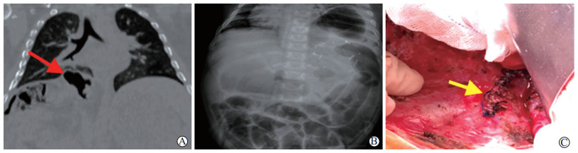
图 1 例1患儿膈疝的影像学表现
注:A图示胸腹联合CT表现为右侧膈疝;B图为术前CT显示膈疝内容物含右肾;C图示膈肌缺损4 cm×3 cm,未见囊腔。
Figure 1. Imaging manifestations of diaphragmatic hernia in case 1 child

图 2 例2患儿膈疝的影像学表现
注:A图为X线胸片显示右侧胸腔充满肠袢,符合右侧膈疝影像表现;B图为胸腹联合CT检查显示膈疝内容物含右肾;C图为常规腹部超声检查提示肝内胆管扩张。
Figure 2. Imaging manifestations of diaphragmatic hernia in case 2 child

图 3 例3患儿膈疝的影像学表现
注:A图为膈疝发生前1 d胸部CT检查显示右侧膈肌薄弱区;B图为X线胸片显示右侧胸腔内充满肠袢伴有典型胃肠道梗阻表现,符合膈疝诊断;C图示一期缝合进行膈肌缺损修补。
Figure 3. Imaging manifestations of diaphragmatic hernia in case 3 child
表 1 3例儿童活体肝移植受者临床资料
Table 1. Clinical data of 3 recipients undergoing pediatric living donor liver transplantation
例序 手术史 PELD②
评分(分)移植物
重量(g)GRWR③
(%)术程
(min)例1 Kasai术① 12 255 3.92 405 例2 Kasai术 8 282 3.03 390 例3 Kasai术 8 245 3.50 430 注:①Kasai术为肝门空肠吻合术。
②PELD为儿童终末期肝病模型。
③GRWR为移植物与受者质量比。 下载: 导出CSV
-
[1] GOLDMAN M, PRANIKOFF T. Biliary disease in children[J]. Curr Gastroenterol Rep, 2011, 13(2):193-201. DOI: 10.1007/s11894-010-0169-1. [2] MOON SB, JUNG SM, KWON CH, et al. Posteromedial diaphragmatic hernia following pediatric liver transplantation[J]. Pediatr Transplant, 2012, 16(4):E106-E109. DOI: 10.1111/j.1399-3046.2010. 01462.x. [3] FORTUNATO AC, PINHEIRO RS, NACIF LS, et al. Hepatic artery thrombosis in liver transplantation in adult recipients using pediatric deceased donors[J]. Transplant Proc, 2020, DOI: 10.1016/j.transproceed.2020.02.034[Epub ahead of print]. [4] LAM HD, MEJIA J, SOLTYS KA, et al. Right diaphragmatic hernia after liver transplant in pediatrics: a case report and review of the literature[J]. Pediatr Transplant, 2013, 17(3):E77-E80. DOI: 10.1111/petr.12052. [5] EARL TM, WELLEN JR, ANDERSON CD, et al. Small bowel obstruction after pediatric liver transplantation: the unusual is the usual[J]. J Am Coll Surg, 2011, 212(1):62-67. DOI: 10.1016/j.jamcollsurg.2010.09.017. [6] OKAJIMA H, HAYASHIDA S, IWASAKI H, et al. Bowel obstruction due to diaphragmatic hernia in an elder child after pediatric liver transplantation[J]. Pediatr Transplant, 2007, 11(3):324-326. http://cn.bing.com/academic/profile?id=2648ba9ec170ba1b16b7b930175823c6&encoded=0&v=paper_preview&mkt=zh-cn [7] HASHMI MU, ULLAH K, TARIQ A, et al. Morgagni-larrey hernia: a possible cause of recurrent lower respiratory tract infections[J]. Cureus, 2019, 11(2):e4035. DOI: 10.7759/cureus.4035. [8] DAO DT, PATEL N, HARTING MT, et al. Early left ventricular dysfunction and severe pulmonary hypertension predict adverse outcomes in "low-risk" congenital diaphragmatic hernia[J]. Pediatr Crit Care Med, 2020, DOI: 10.1097/PCC.0000000000002318[Epub ahead of print]. [9] OKUR MH, YANKOL Y, MECIT N, et al. Diaphragmatic hernia after liver transplant in children: report of 2 cases[J]. Exp Clin Transplant, 2018, 16(3):337-339. DOI: 10.6002/ect.2015.0242. [10] EMAMAULLEE JA, NEKRASOV V, GILMOUR S, et al. Case series and systematic review of acquired diaphragmatic hernia after liver transplantation[J]. Pediatr Transplant, 2018, 22(8):e13296. DOI: 10.1111/petr.13296. [11] WANG K, GAO W, MA N, et al. Acquired diaphragmatic hernia in pediatrics after living donor liver transplantation: three cases report and review of literature[J]. Medicine (Baltimore), 2018, 97(15):e0346. DOI: 10.1097/MD.0000000000010346. [12] COVARRUBIAS K, LUO X, MASSIE A, et al. Determinants of length of stay after pediatric liver transplantation[J]. Pediatr Transplant, 2020:e13702. DOI: 10.1111/petr.13702. [13] SUZUKI K, KOMURA M, TERAWAKI K, et al. Engineering and repair of diaphragm using biosheet (a collagenous connective tissue membrane) in rabbits[J]. J Pediatr Surg, 2018, 53(2):330-334. DOI: 10.1016/j.jpedsurg.2017.11.035. [14] TAGUCHI T, YANAGI Y, YOSHIMARU K, et al. Regenerative medicine using stem cells from human exfoliated deciduous teeth (SHED): a promising new treatment in pediatric surgery[J]. Surg Today, 2019, 49(4):316-322. DOI: 10.1007/s00595-019-01783-z. [15] QU W, ZHU Z, WEI L, et al. Paediatric liver re-transplantation after primary partial liver graft transplantation: a report of four cases[J]. Int J Clin Pract, 2016, 70 (Suppl 185):31-34. DOI: 10.1111/ijcp.12814. [16] DÖKÜMCÜ Z, DIVARCI E, ERDENER A, et al. Acquired right diaphragmatic hernia following pediatric living donor orthotopic liver transplantation[J]. Pediatr Transplant, 2015, 19(6):E149-E151. DOI: 10.1111/petr.12548. [17] EKSER B, MANGUS RS. Spontaneous bacterial peritonitis[J]. Lancet, 2017, 389(10070):735. DOI: 10.1016/S0140-6736(16)30782-6. [18] TRAN LT, CARULLO PC, BANH DPT, et al. Pediatric liver transplantation: then and now[J]. J Cardiothorac Vasc Anesth, 2020, DOI: 10.1053/j.jvca.2020.02.019[Epub ahead of print]. [19] ESPOSITO F, LIM C, SALLOUM C, et al. Diaphragmatic hernia following liver resection: case series and review of the literature[J]. Ann Hepatobiliary Pancreat Surg, 2017, 21(3):114-121. DOI: 10.14701/ahbps.2017.21.3.114. [20] GARNON J, CAZZATO RL, AULOGE P, et al. Adjunctive hydrodissection of the bare area of liver during percutaneous thermal ablation of sub-cardiac hepatic tumours[J]. Abdom Radiol (NY), 2020, DOI: 10.1007/s00261-020-02463-0[Epub ahead of print]. [21] LOCHAN R, SAIF R, GANJOO N, et al. Diaphragmatic herniation following donor hepatectomy for living donor liver transplantation: a serious complication not given due recognition[J]. Ann Hepatobiliary Pancreat Surg, 2017, 21(4):232-236. DOI: 10.14701/ahbps.2017.21.4.232. [22] OH JW, OH SN, JUNG SE, et al. Diaphragmatic hernia after living-donor right hepatectomy: an important late donor complication[J]. J Comput Assist Tomogr, 2017, 41(5):726-730. DOI: 10.1097/RCT.0000000000000591. [23] MCCABE AJ, ORR JD, SHARIF K, et al. Right-sided diaphragmatic hernia in infants after liver transplantation[J]. J Pediatr Surg, 2005, 40(7):1181-1184. doi: 10.1016/j.jpedsurg.2005.03.063 [24] HEFFRON T, WELCH D, PILLEN T, et al. Liver transplant in a four-month-old child with biliary atresia, unilateral pulmonary agenesis, and diaphragmatic hernia: first case report[J]. Pediatr Transplant, 2006, 10(4):513-516. http://search.ebscohost.com/login.aspx?direct=true&db=aph&AN=20857834&site=ehost-live [25] YAZDANI M, PARK E, UDAYASANKAR U. Postsurgical diaphragmatic herniation: a rare delayed complication of pediatric intraabdominal surgery[J]. J Pediatr Surg, 2016, 51(2):333-335. DOI: 10.1016/j.jpedsurg.2015.11.019. [26] TESTINI M, GIRARDI A, ISERNIA RM, et al. Emergency surgery due to diaphragmatic hernia: case series and review[J]. World J Emerg Surg, 2017, 12:23. DOI: 10.1186/s13017-017-0134-5. -

 下载:
下载:


 点击查看大图

计量
- 文章访问数: 237
- HTML全文浏览量: 221
- PDF下载量: 26
- 被引次数: 0